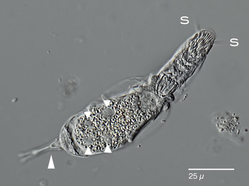
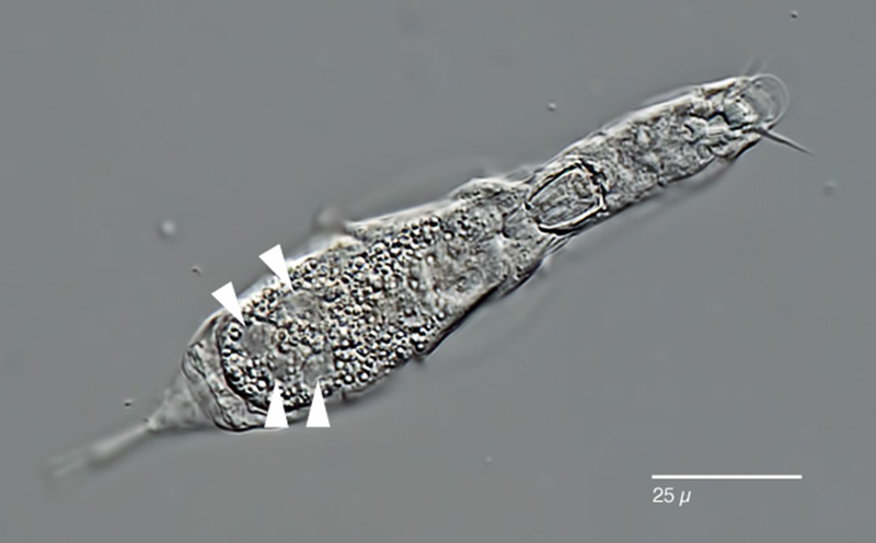
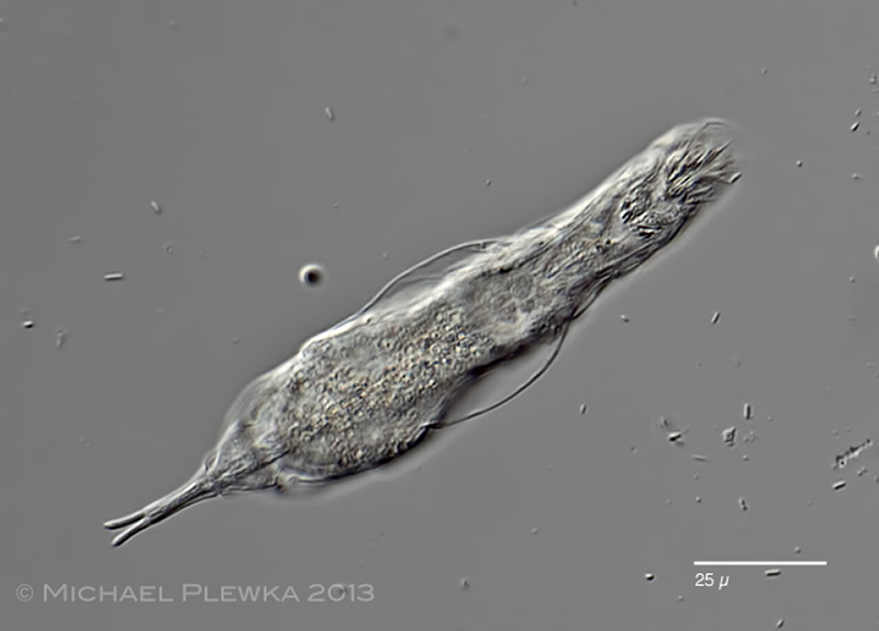
| Bryceella perpusilla; very small (body length: 100µm), extremely vivid species; ventral view showing the ciliary field of the head. The identification is based on the apical styli (S) and the vitellarium with 4 nuclei (triangles). The arrowhead points to the caudal antenna.(1) |
| |
 |
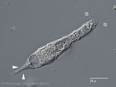
| Bryceella perpusilla; arrowheads pointing to the 4 nuclei of the vitellarium. (1) |
| |
 |
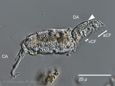
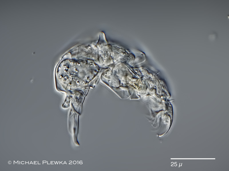
| Bryceella perpusilla; focus on the outwardly curved toes, which can be retracted like a telescope into the last pseudosegment of the foot. (1) |
| |
 |
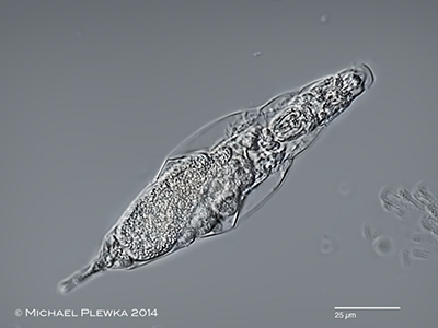
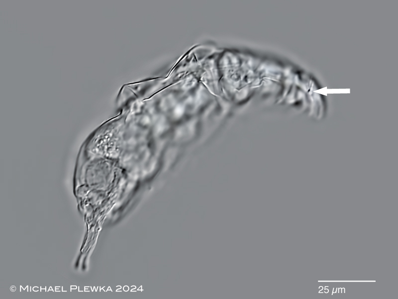
| Bryceella perpusilla; lateral view; specimen from (3). |
| |
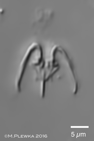 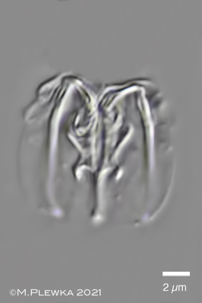 |
| Bryceella perpusilla; modified malleate trophi; left: specimen from (3); right: specimen from (4). |
| |
 |
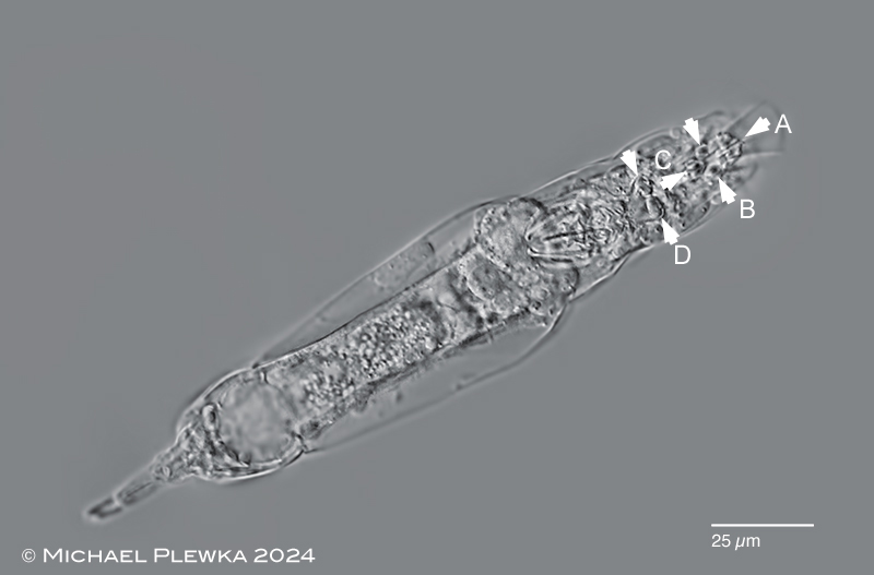 |
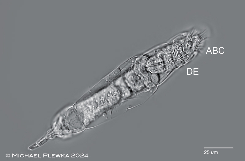
Bryceella perpusilla; dorsoventral view of a young specimen. This is a still image of a YT-video that shows the movement of the cilia of the corona.
Upper image: the ventral cilia (?bristles?) are organised in 8 groups which insert at 8 hypodermal cushions (6 of them are marked by arrowheads: 1x A; 2x B; 1x C; 2x D in the lower image). The cilia bundles A, B, C appear to be primarily responsibe for locomotion. The cilia of group DE are separated from the other group by a membrane and are involved in feeding. (5) |
| |
 |
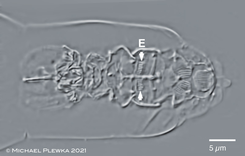 |
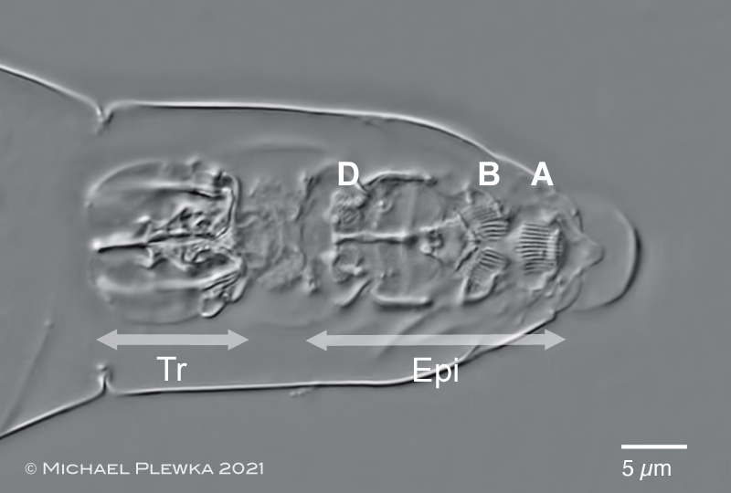
| Bryceella perpusilla; two images of the anterior part of a specimen macerated with SDS in dorsoventral view. Upper image: some epipharyngeal structures ("Epi", marked by double arrow) can be observed aside from the trophi (Tr). The striated structures A, B, D are the base of the ciliary areas shown in the above images. Lower image: slightly different focal plane shows another group of striated structures ( arrowheads: E) (4). |
| |
 |
 |
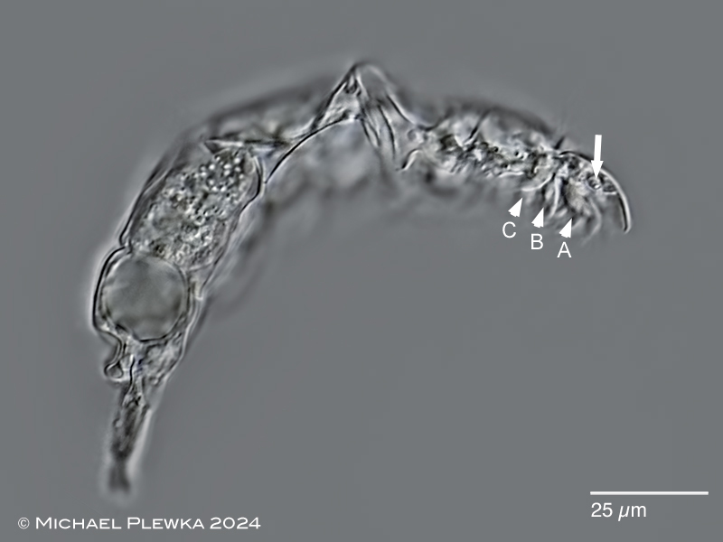
| Bryceella perpusilla; lateral view; upper image: the arrowheads point to some groups (A; B; C) of the ventral cilia/ ?bristles?. Cilia group D is not in focus of these images. The arrow points to the base of the right of the ?sensory? bristles / "styli" which are in permanent movement. lower image: different focal plane; arrow marks the right stylus.(5) |
| |
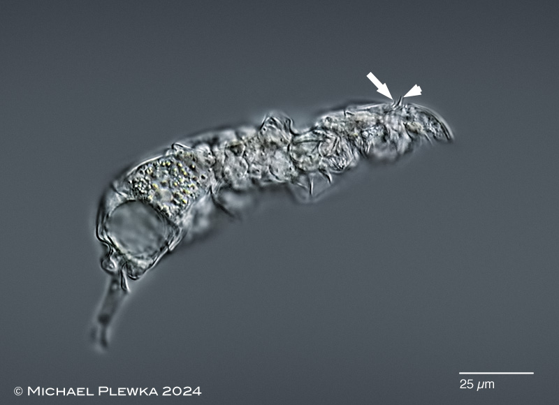 |
| Bryceella perpusilla; lateral view; focal plane on the epidermal projection (arrowhead) and the dorsal antenna (arrow). (5) |
| |
|
| Bryceella perpusilla; upper left: specimen from (1); dorsoventral view. Focus on styli and caudal antennae. Upper right: lateral view of specimen from (7). Lower left: specimen from (2). |
| |
| It might be possible that the species identified as Wierzejskiella vagneri (earlier found in nature reserve Schwalm) is also Bryceella perpusilla. |
| |
| Sample (4) courtesy of Dr. D. Fontaneto, Verbania, Italy |
| |
| |
|
|
| |
| |
|
|
| |
| |
|
|
| |
| |
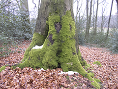
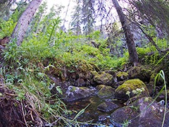
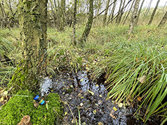
| Location: Hattingen Oberstueter, forest (1); (2); Arendal, Norway (4); Felderbachtal, Hattingen, NRW, Germany (7) |
| Habitat: tree moss, sympatric with B.stylata (1); (2); sphagnum moss (3); (4); between the liverwort Metzgeria sp. s(7) |
| Date: ; 31.8.2014 (2); 04.08.2016 (3); 27.10.2021 (4); 03.03. 2015 (7) |
|
| |
| |
|
|